Diffusion MRI (DMRI) is an emerging non-invasive medical imaging technique primarily used to study white matter structures in brain. Based on the principles of Nuclear Magnetic Resonance (NMR), it utilizes the fact that diffusion of water molecules in
white matter region is not isotropic. Presence of nerve fibers, membranes and other macro-molecules creates a diffusion pattern that can be used to study microscopic details and fiber tracts. In DMRI, signals are captured along many diffusion
directions that is a time intensive process. Ideally, patients are required to remain stationary during this time while the data is being captured and is very inconvenient. At SBILab, we are working on accelerated reconstruction of DMRI,
particularly, HARDI signals using compressive sensing methods via collecting fewer data. The CS-based reconstructed signal is aimed to achieve comparable DMRI representations like ODF and FA with lesser signal samples, thereby decreasing
the scanning time. In the near future, we would be working on fibre tractography in HARDI.
MSR-HARDI method : This is our recently proposed compressive sensing based reconstruction method using predefined seperable dictionaries.
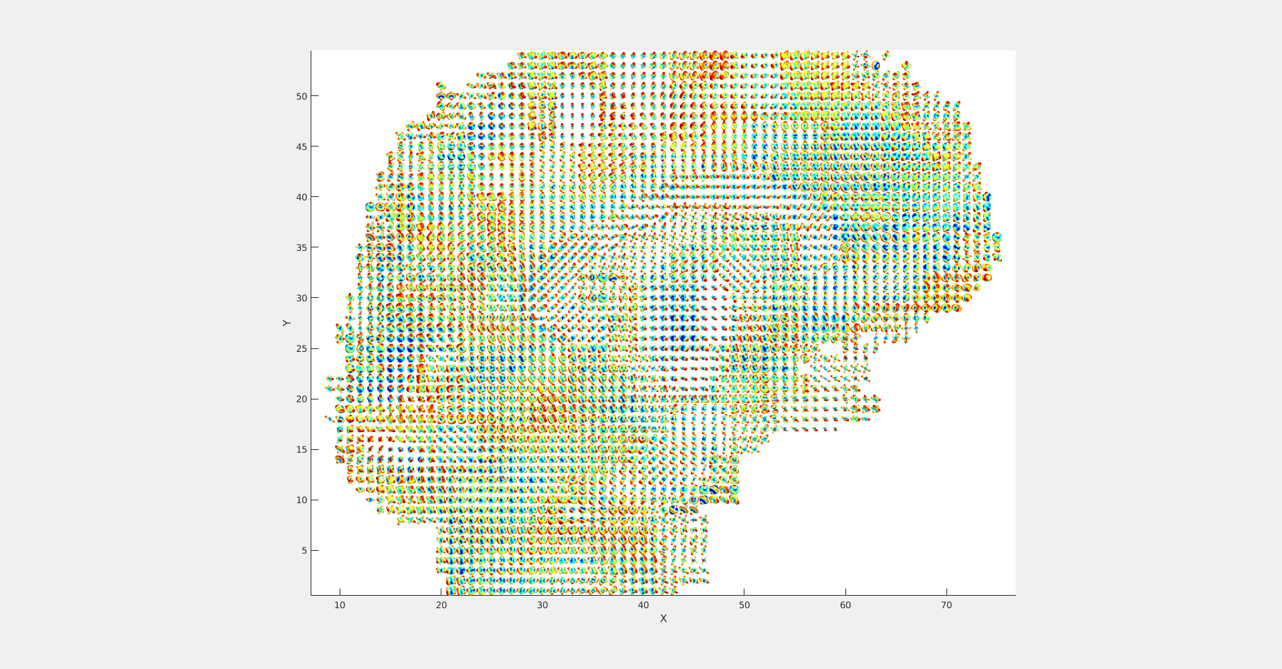
MSR-HARDI: Orientation Distribution Functions (ODFs) depicting white matter nerve fiber directions in corpus callosum region. ODFs are generated over image reconstructed using MSR-HARDI method with only
1% (k-q) space samples (i.e. 10% k-space and 10% qspace samples).